Diagram Of Hip.and Back.muscles / Ligaments Tendons And Muscles Of The Hip Joint Naples Best Hip Surgeon. The back muscles represented on an anatomical chart and on a schematic view of the origin and insertion of the proper muscles of the back (iliocostal muscle of the neck, lumbar (lumbar and thoracic parts), longissimus muscles of head, neck and thorax, the spinalis muscles of the neck and thorax, semispinalis muscle of the head, neck and thorax. Piriformis, external and internal obturators and the superior and inferior gemelli. Causes of tightness a couple of the most obvious causes for muscle tightness in your hips and lower back are acute injuries — such as muscle strains — or simple soreness from doing more exercise than your body. Back muscle diagrams labeled 12 photos of the back muscle diagrams labeled back muscle diagrams labeled, lower back muscle diagrams labeled, human muscles, back muscle diagrams labeled, lower back muscle diagrams labeled. This is a diagram of the larger and more surface muscles of the low back.
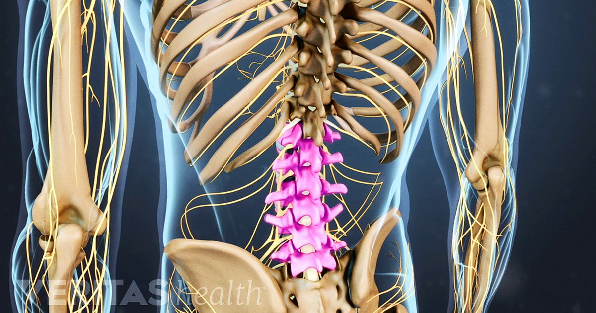
The muscles of the lower back help stabilize, rotate, flex, and extend the spinal column, which is a bony tower of 24 vertebrae that gives the body structure and houses the spinal cord. It runs from your lower back through your pelvis, passing to the front of your hip where it attaches to the top of your femur, which is your thigh bone. The pubis, ischium, and ilium together constitute the pelvis while the thigh bone is the femur. The hip muscles are going to be slip into hip muscles and gluteal muscles. Nerves carry signals from the brain to the muscles to move the hip and carry signals from the muscles back to the brain about pain, pressure and temperature.
Some Reasons Why You Should Stop Stretching Your Hip Flexors Deansomerset Com from deansomerset.com It is the most superficial of all the back muscles. Hip anatomy diagram from bones to joints science trends. Lower back muscle diagram anatomy Like the forearm, the upper leg, or thigh, has a dense arrangement of many muscles. The hip abductors consist of the: This is a diagram of the larger and more surface muscles of the low back. The muscles on each side form a trapezoid shape. The part of the nerve that emerges out of the spine is called the nerve root.
The hip joint is made up of two.
On the anterior side, the most prominent of the muscles are the sartorius muscle and the four muscles that make up quadriceps muscle group (the quads.) Back muscle diagrams labeled 12 photos of the back muscle diagrams labeled back muscle diagrams labeled, lower back muscle diagrams labeled, human muscles, back muscle diagrams labeled, lower back muscle diagrams labeled. Causes of tightness a couple of the most obvious causes for muscle tightness in your hips and lower back are acute injuries — such as muscle strains — or simple soreness from doing more exercise than your body. The bones together make up the hip. The muscles of the lower back help stabilize, rotate, flex, and extend the spinal column, which is a bony tower of 24 vertebrae that gives the body structure and houses the spinal cord. An important group of muscles in the pelvis is the pelvic floor. The fibres attach to the clavicle, acromion and the scapula spine. In physical therapy, a therapist will determine if you need to stretch the lower back muscles and other muscles such as the piriformis or hamstrings. These muscles can be grouped based upon their location and function. To learn more about the anatomy of the spine, watch this video. The back is the body region between the neck and the gluteal regions. 1 hip anatomy, function and common problems. Related posts of muscles of the lower back and hip diagram muscle anatomy neck.
The trapezius is a broad, flat and triangular muscle. An important group of muscles in the pelvis is the pelvic floor. These muscles can be grouped based upon their location and function. The extensor muscles are attached to back of the spine and enable standing and lifting objects. Muscle anatomy neck 12 photos of the muscle anatomy neck dog neck muscle anatomy, front neck muscle anatomy, muscle anatomy neck, muscle anatomy of neck and shoulder, neck muscle anatomy chart, human muscles, dog neck muscle anatomy, front neck muscle anatomy, muscle anatomy neck, muscle anatomy of neck and.
Understanding Lower Back Anatomy from embed.widencdn.net See back muscles and low back pain. The many muscles of the hip provide movement, strength, and stability to the hip joint and the bones of the hip and thigh. Browse our library of free human anatomy images and pictures. An important group of muscles in the pelvis is the pelvic floor. The bones together make up the hip. The hip joint is made up of two. In a nutshell, tight outer hip muscles, also known as. In physical therapy, a therapist will determine if you need to stretch the lower back muscles and other muscles such as the piriformis or hamstrings.
1 hip anatomy, function and common problems.
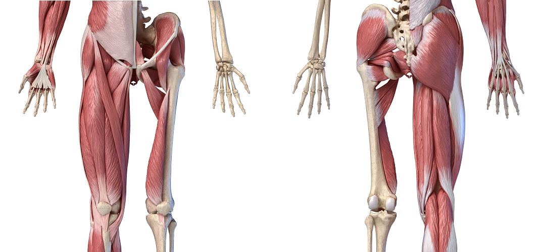
This video also provides you with a. Common causes of tight hip and lower back muscles include injury, too little activity, too much activity and muscular imbalances. The muscles of the lower back help stabilize, rotate, flex, and extend the spinal column, which is a bony tower of 24 vertebrae that gives the body structure and houses the spinal cord. If you are starting to feel hip pain or stiffness, you'll want to know more about the bones and muscles that make up the hip's anatomy. As you can see from the diagram to the right, there are many muscles and tendons that make up the hip and buttocks region. Hip anatomy diagram from bones to joints science trends. Like the forearm, the upper leg, or thigh, has a dense arrangement of many muscles. It runs from your lower back through your pelvis, passing to the front of your hip where it attaches to the top of your femur, which is your thigh bone. The fibres attach to the clavicle, acromion and the scapula spine. Related posts of muscles of the lower back and hip diagram muscle anatomy neck. The back muscles stabilize your spine. The anatomy of the fascia lata and iliotibial tract. Muscles located at the side of the hip, which include the gluteus medius, piriformis, and hip external rotator muscles contribute greatly to the well being of your lower back, as well as your posture.when these muscles get tight, as they often do, you may find that along with hip pain, your lower back hurts—but you can't figure out why.
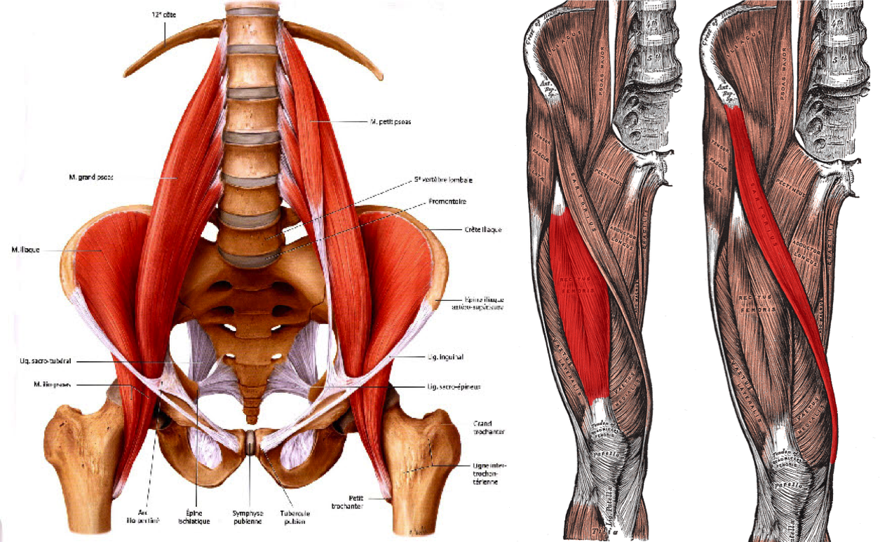
Most of the time, back muscle pain is diagnosed then treated with little more than a prescription of rest, painkillers and muscle relaxants. Three types of back muscles that help the spine function are extensors, flexors and obliques. The psoas major is a large muscle that runs from the bodies and disc of the l1 to l5 vertebrae, joins with the iliacus via its tendon, and connects to the lesser trochanter of the femur. In physical therapy, a therapist will determine if you need to stretch the lower back muscles and other muscles such as the piriformis or hamstrings. Lower back muscle diagram anatomy
Hip Muscles The Definitive Guide Biology Dictionary from biologydictionary.net The many muscles of the hip provide movement, strength, and stability to the hip joint and the bones of the hip and thigh. The four groups are the anterior group, the posterior group, adductor group, and finally the abductor group. The part of the nerve that emerges out of the spine is called the nerve root. To learn more about the anatomy of the spine, watch this video. Back muscle diagrams labeled 12 photos of the back muscle diagrams labeled back muscle diagrams labeled, lower back muscle diagrams labeled, human muscles, back muscle diagrams labeled, lower back muscle diagrams labeled. The bones of the hip include the femur, the ilium, the ischium, and the pubis. This is a diagram of the larger and more surface muscles of the low back. The muscles of the abdomen, lower back, and pelvis are separated from those of the chest by the muscular wall of the diaphragm, the critical breathing muscle.
Diagram demonstrating the posterior view of the piriformis muscle orientation, origin and insertion on the pelvis and femur.
The bones together make up the hip. Hip anatomy diagram from bones to joints science trends. The anatomy of the fascia lata and iliotibial tract. The trapezius is a broad, flat and triangular muscle. To learn more about the anatomy of the spine, watch this video. The iliacus originates on the iliac fossa of the ilium. The muscles of the lower back help stabilize, rotate, flex, and extend the spinal column, which is a bony tower of 24 vertebrae that gives the body structure and houses the spinal cord. The back muscles represented on an anatomical chart and on a schematic view of the origin and insertion of the proper muscles of the back (iliocostal muscle of the neck, lumbar (lumbar and thoracic parts), longissimus muscles of head, neck and thorax, the spinalis muscles of the neck and thorax, semispinalis muscle of the head, neck and thorax. The hip joint is made up of two. The fibres attach to the clavicle, acromion and the scapula spine. Muscles of the gluteal region. It runs from your lower back through your pelvis, passing to the front of your hip where it attaches to the top of your femur, which is your thigh bone. In physical therapy, a therapist will determine if you need to stretch the lower back muscles and other muscles such as the piriformis or hamstrings.